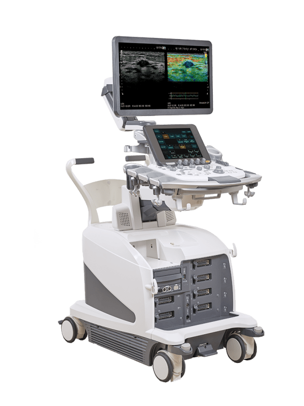
During a lifetime, women need access to various screening options. Ranging from pregnancy checks, to fetal well-being, and oncological care of female organs like uterus, ovaries, and breast – Fujifilm tailors comprehensive solutions that support women through all stages of life.
From Early to Late Pregnancy Stages
- Comprehensive Fetal Assessment
- Fetal Heart Solutions
- 3D/4D Imaging
From Early Screening to Treatment Plans
- Gynaecological Imaging
- Infertility Treatment
- Breast Imaging
From Early To Late Pregnancy Stages
Image Optimization
The combination of our image processing, specialized probes, and superior image optimization work together to capture the subtlest of signals. Rest assure of crystal-clear, high resolution images for confident assessment of fetal well-being, and easy explanation of results to new parents.
Dynamically Focused Images

Our eFocusing technology provides auto-optimization of images from near to far field, without compromise on frame rate. Delivering homogeneous and sharp B-mode images for your accurate assessment.
Highly Sensitive Colour Flow

Spot micro-vasculature that you’ve never been able to see before, with our eFlow and Detective Flow Imaging modes! The higher sensitivity allows for easy visualization of very slow blood flow, or that in tiny vessels. Additionally, DFI removes artefacts from the actual blood flow to deliver highly accurate information on blood perfusion.
For That Added 3D-Gloss

For a 3D-like look, our Glossy mode adds vivid highlights on the blood flow. This helps improve visibility and simplifies your understanding of the structural anatomy, flow patterns and positional relationship of vessels.
Streamlined Examinations
Your data is just one click away with our AI-based automatic measurements for fetal biometry and the estimated fetal weight. What more you can calculate the nuchal translucency, fetal heart rate and fractional shortening – all automatically. So when it comes to speeding up your exams, increasing accuracy and reducing your own stress… just let the system take the strain.
Auto-Biometry

The four major fetal structures (BPD, HC, AC and FL) required for biometric measurement is automatically measured via a one touch operation.
Auto Fetal Heart Measurements

To help improve detection rate, we offer a sophisticated fetal heart imaging package that includes automated measurement of important metrices like heart rate and fractional shortening so you get reliable and reproducible results at all times.
One Probe Solution

Lightweight and ergonmically designed, our OB-dedicated volume probe incorporates the highest resolution, penetration, and a superwide frequency bandwidth that enables 2D to 4D screening, from early all the way to late pregnancy stages.
Fetal Heart Solutions
Congenital heart disease in infants is a challenging issue with structural cardiac anomalies often missed by prenatal ultrasound. To help improve detection rate, we offer a sophisticated fetal heart imaging package that goes beyond screening to give you clear visibility and valuable insights you need to deliver better patient outcomes.
Auto Fetal Heart Rate

For all 3 trimesters, AutoFHR+ allows the automatic calculation of fetal heart rate in real-time, offering a safer and more objective measurement compared to conventional Doppler or M-mode methods.
Auto Fractional Shortening

AutoFS automatically tracks fetal heart movement by following the displacement of the heart wall to assess fetal heart contractility. This approach delivers precise accuracy as the measurement is unaffected by change in fetal position or by mother’s breathing.
Dynamic Slow-Motion Display

To better observe and analyse the fast moving heart, we support a dynamic display of both the real-time view and its slow-motion counterpart, simultaneously, side-by-side.
Dual Gate Doppler

With DGD, you can compare 2 waveforms from different locations – in the same heart cycle, giving you stable doppler measurements to assess cardiac function and conditions like fetal arrhythmia with ease and reliability.
Convex Continuous Wave

Our convex transducer is capable of supporting Continuous Wave mode for sampling high velocity blood flow that exceed the capability of Pulsed Wave, useful for cases like valvular regurgitation, vessel stenosis etc. All of which can be done without having to change transducers.
Spatio-Temporal Image Correlation

For the fast-moving fetal heart, STIC allows for multi-slice 3D volume data sets of one cardiac cycle to be reconstructed for a better understanding of the fetal heart.
3D Evaluation

With images obtained in STIC, you can reconstruct heartbeats in any cross section or angle for detailed analysis in 3D view.
3D visualization makes it easier to evaluate the positioning of arteries, aortic arch, or pattern of the aorta to determine any abnormally present.
Myocardial Function Analysis

Get advanced quantitative measurements of the cardiac wall movement with our 2DTT speckle tracking technology. You can assess various parameters for strain, volume and thickness: like Global Longitudinal Strain, cardiac structure, myocardial contractility, torsion/displacement, and wall thickening.
Tissue Doppler Imaging

Employ doppler measurements in cardiac muscle walls instead of blood flow with TDI. You can get a deeper insight into the cardiac cycle with comprehensive information like myocardial velocity, displacement, or deformation.
3D/4D Imaging
Three- and four-dimensional imaging delivers photo-realistic images that can strengthen maternal-fetal bonding, as well as adding important clinical information to grasp complicated conditions, and give a thorough assessment of any deformation.
Realistic Vue

Adding 3D depth with shadows, highlights and liquid effects can significantly improve the visibility of any small anomalies like cleft lips, skill deformation, club feet or polydactyly.
Our AutoClipper also quickens the process by automatically defining the optimal cut plane, removing placental and other unwanted tissue signals in front of the fetus to reveal a beautiful fetal image
4DShading Flow

You can now go one step deeper in evaluating vascular information with 3D/4D effect. This enables us to grasp complicated conditions such as TAPVC, and detect abnormal blood flow in the fetal brain, or umbilical cord.
Curved MPR

Neural tube defects, open spina bifida, or a partly missing corpus callosum are common central nervous system malformations which require high ultrasound expertise to examine. Our CMPR mode makes it easier: you can render a 3D image of curved anomalies and obtain any plane from it by simply drawing a line.
4D Translucence

Apply translucence on your ultrasound image to see beyond the surface of the body into developing internal anatomy. Organ boundaries, vessel walls, brain cavities, gastrointestinal anatomy, or even liquid structures in early embryo brain can be observed for an accurate assessment.
From Early Screening To Treatment Plans
Use 3D Imaging to improve visualization of the uterus and ovarian anatomy
Understanding uterine cavities and anatomy or ovarian structures becomes an easier task with added 3D depth, shadows, highlights, glossy effects and translucency to boost the visibility. 3D also allows you to look at structures in the coronal plane, which isn’t possible with just 2D sonography.

Easily display curved structures in the best plane for easy assessment
Curved anatomical structures need high ultrasound expertise to get the full picture – which is why we designed the CMPR mode. You can now display a 3D coronal image of curved anatomies, like the uterus, and obtain your desired plane by simply drawling a line.

Perfect your Oncological Treatment with Fusion Imaging that helps you localise, classify, and perform biopsies with the highest precision.
Ultrasound is a quick, easy and affordable tool for managing gynaecological conditions like ovary/cervical tumours, cysts, endometriosis, or adenomyosis.
Benefit from multiparametric imaging with our Real-time Virtual Sonography that fuses real-time ultrasound image with MRI, CT and PET data to give you a deeper understanding of your patient’s condition and support you in performing treatment plans with the highest accuracy.

Conduct infertility treatment with the right tools for the best patient outcome
With our slim 200° FOV angled transvaginal transducer, you can obtain clear images of the ovaries, endometrial lining, uterus, or fallopian tubes with minimal manipulation and extra comfort for patients.
The probe also supports biopsy exams by guiding the biopsy needle during in-vitro fertilization.

Supported by Multi-Follicle Volume to speed up the process

Multi-follicle Volume helps to automatically measure up to 20 follicles in the ovary using the volume data.
And Trace Volume Measurement to accurately determine the timing of implanting the fertilized ovum

Trace Volume Measurement helps measure the volume of uterine endometrium accurately, providing important information to determine the rate of implantation of fertilized ovum for a successful result.
Tools for Easy Visualization
To detect early signs of abnormalities, our ultrasound system and transducers are optimized to deliver the purest of images, and helpful features to make accurate diagnosis, and speedy breast examinations.
Carving mode to characterize the smallest abnormaly at the earliest stage

The premium Carving Imaging tool gives you a clear delineation of tissue structures for a better understanding of what you’re dealing with. You can detect shadows around lesions, check if it’s liquid or solid, or confidently guide a cytology needle for biopsy.
Real-time Tissue Elasticity makes it easy to differentiate between benign and malignant lesions

RTE helps you better visualize tissue elasticity by displaying the stiffness as a colour map, which can be interpreted according to the Tsukuba or BI-RADS scale. Quantitatively, our system will also automatically identify the region of interest, so you can calculate strain ratio or fat-lesion ratio with a single click.
See all layers of the breast all at once with our unique CMUT transducer

Save yourself the hassle and costs of changing probes with our wideband CMUT probe. Regardless of the size, thickness or density of the mammary gland, you can view the entire breast – from the skin to the pleura, ribs, and thoracic wall in perfect clear detail. Even difficult breasts (like type IV density, post-operative, or large volume) are easy to scan with CMUT.
Tools for Precise and Successful Procedures
For the best treatment outcomes, we support you with tools that help you plan out the best procedures, and guidance to perform surgery with the highest accuracy.
Vascular Imaging

Guide your treatment steps by assessing vascularity both inside and around suspicious areas – including the viewing of tiny and slow vessels with our eFlow and Detective Flow Imaging modes. Flow patterns can also be monitored with closer detail by pairing with Contrast Imaging (before, during, or after treatment).
Fusion imaging

Compare your patient’s MRI images with real-time ultrasound to get a complete, conclusive confirmation of the lesion location. You can plan preoperative markings or needle paths; increase precision in interventional procedures; and monitor treatment success after chemical therapy.
Guided Markings

Our transducers come with physical markings on the head so you can easily reference and paint mark-ups onto the breast skin prior to surgery. These lines correspond to digital lines displayed on B-mode image, which serves as a useful positional guide to understand which area you are working with between the organ and the probe.


